Biological Image Analysis
144 items from hallvaaw/awesome-biological-image-analysis ★198
-
Classpose github.com
A foundation model-driven whole slide image-scale cell phenotyping method with QuPath integration.
-
Napari-aicsimageio github.com
Multiple file format reading directly into napari using pure Python.
-
PartSeg github.com
A GUI and a library for segmentation algorithms.
-
Cellshape github.com
3D single-cell shape analysis of cancer cells using geometric deep learning.
-
HistoClean github.com
Tool for the preprocessing and augmentation of images used in deep learning models.
-
LLSpy github.com
Python library to facilitate lattice light sheet data processing.
-
Quanfima github.com
Quantitative Analysis of Fibrous Materials: A collection of useful functions for morphological analysis and visualization of 2D/3D data from various areas of material science.
-
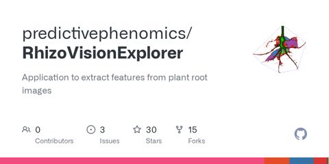
Rhizovision Explorer github.com
Free and open-source software developed for estimating root traits from images acquired from a flatbed scanner or camera.
-
BoneJ github.com
Collection of Fiji/ImageJ plug-ins for skeletal biology.
-
YeaZ github.com
An interactive tool for segmenting yeast cells using deep learning.
-
NCS github.com
Noise correction algorithm for sCMOS cameras.
-
ColiCoords github.com
Python project for analysis of fluorescence microscopy data from rodlike cells.
-
Ijp-color github.com
Plugins for ImageJ - color space conversions and color calibration.
-
Flika github.com
An interactive image processing program for biologists written in Python.
-
Z-stack Depth Color Code github.com
ImageJ/Fiji plugin to colorcode Z-stacks/hyperstacks.
-
SyMBac github.com
Accurate segmentation of bacterial microscope images using synthetically generated image data.
-
ASI_MTF github.com
ImageJ macro to calculate the modulation transfer function (MTF) based on a knife edge (or slanted edge) measurement.
-
TrackMateR github.com
R package to analyze cell migration and particle tracking experiments using outputs from TrackMate.
-
MIA github.com
Fiji plugin which provides a modular framework for assembling image and object analysis workflows.
-
Em-scalebartools github.com
Fiji/ImageJ macros to quickly add a scale bar to an (electron microscopy) image.
-
Fungal Feature Tracker (FFT) github.com
Tool to quantitatively characterize morphology and growth of filamentous fungi.
-
CompactionAnalyzer github.com
Python package to quantify the tissue compaction (as a measure of the contractile strength) generated by cells or multicellular spheroids that are embedded in fiber materials.
-
ZVQ - Zebrafish Vascular Quantification github.com
Image analysis pipeline to perform 3D quantification of the total or regional zebrafish brain vasculature using the image analysis software Fiji.
-
Cytominer-database github.com
Command-line tools for organizing measurements extracted from images.
-
CellMigration github.com
Analysis of 2D cell migration in Igor.
-
DetecDiv github.com
Comprehensive set of tools to analyze time microscopy images using deep learning methods.
-
QuimP github.com
Software for tracking cellular shape changes and dynamic distributions of fluorescent reporters at the cell membrane.
-
XitoSBML github.com
ImageJ plugin which creates a Spatial SBML model from segmented images.
-
htsimaging github.com
Python package for high-throughput single-cell imaging analysis.
-
Empanada github.com
Panoptic segmentation algorithms for 2D and 3D electron microscopy images.
-
Salem² github.com
Segment Anything in Light and Electron Microscopy via Membrane Guidance.
-
BactMap github.com
A command-line based R package that allows researchers to transform cell segmentation and spot detection data generated by different programs into various plots.
-
PhenotyperCV github.com
Header-only C++11 library using OpenCV for high-throughput image-based plant phenotyping.
-
DypFISH github.com
Python library for spatial analysis of smFISH images.
-
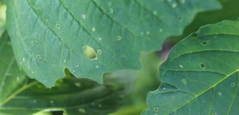
LeafByte zoegp.science
Free and open source mobile app for measuring herbivory quickly and accurately.
-
YeastMate yeastmate.readthedocs.io
Neural network-assisted segmentation of mating and budding events in S. cerevisiae.
-
VisionSetil visionsetil.com
Multi-view image classification of Iberian mushrooms for education (open encyclopedia of 523 taxa, deadly lookalikes). Orientation only — never consumption permission.
-
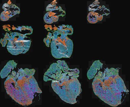
TissUUmaps tissuumaps.github.io
Visualizer of NGS data, plot millions of points and interact, gate, export. ISS rounds and base visualization.
-
Trackpy soft-matter.github.io
Fast and Flexible Particle-Tracking Toolkit.
-
QuPath qupath.github.io
Open source software for digital pathology image analysis.
-
RhizoTrak prbio-hub.github.io
Open source tool for flexible and efficient manual annotation of complex time-series minirhizotron images.
-
Orbit orbit.bio
A versatile image analysis software for biological image-based quantification using machine learning, especially for whole slide imaging.
-
Large-scale image-based screening and profiling of cellular phenotypes onlinelibrary.wiley.com
A workflow for phenotype extraction from high throughput imaging experiments.
-
Panda nitrc.org
Pipeline for Analyzing braiN Diffusion imAges: A MATLAB toolbox for pipeline processing of diffusion MRI images.
-
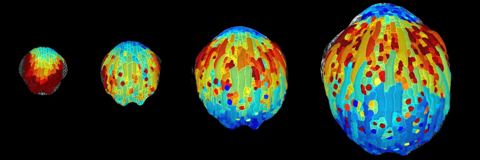
MorphoGraphX morphographx.org
Open source application for the visualization and analysis of 4D biological datasets.
-
PaCeQuant mitobo.informatik.uni-halle.de
An ImageJ-based tool which provides a fully automatic image analysis workflow for PC shape quantification.
-
Biobeam maweigert.github.io
Open source software package that is designed to provide fast methods for in-silico optical experiments with an emphasize on image formation in biological tissues.
-
Workflow and metrics for image quality control in large-scale high-content screens linkinghub.elsevier.com
Approaches for quality control in high-content imaging screens.
-
NeuronJ imagescience.org
An ImageJ plugin for neurite tracing and analysis.
-
TraJClassifier imagej.net
Fiji plugin that loads trajectories from TrackMate, characterizes them using TraJ and classifiies them into normal diffusion, subdiffusion, confined diffusion and directed/active motion by a random forest approach (through Renjin).
- next page of items loading…