Biological Image Analysis
144 items from hallvaaw/awesome-biological-image-analysis ★198
-
Classpose github.com
A foundation model-driven whole slide image-scale cell phenotyping method with QuPath integration.
-
Napari-aicsimageio github.com
Multiple file format reading directly into napari using pure Python.
-
PartSeg github.com
A GUI and a library for segmentation algorithms.
-
Cellshape github.com
3D single-cell shape analysis of cancer cells using geometric deep learning.
-
HistoClean github.com
Tool for the preprocessing and augmentation of images used in deep learning models.
-
LLSpy github.com
Python library to facilitate lattice light sheet data processing.
-
Quanfima github.com
Quantitative Analysis of Fibrous Materials: A collection of useful functions for morphological analysis and visualization of 2D/3D data from various areas of material science.
-
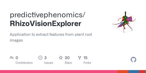
Rhizovision Explorer github.com
Free and open-source software developed for estimating root traits from images acquired from a flatbed scanner or camera.
-
BoneJ github.com
Collection of Fiji/ImageJ plug-ins for skeletal biology.
-
YeaZ github.com
An interactive tool for segmenting yeast cells using deep learning.
-
NCS github.com
Noise correction algorithm for sCMOS cameras.
-
ColiCoords github.com
Python project for analysis of fluorescence microscopy data from rodlike cells.
-
Ijp-color github.com
Plugins for ImageJ - color space conversions and color calibration.
-
Flika github.com
An interactive image processing program for biologists written in Python.
-
Z-stack Depth Color Code github.com
ImageJ/Fiji plugin to colorcode Z-stacks/hyperstacks.
-
SyMBac github.com
Accurate segmentation of bacterial microscope images using synthetically generated image data.
-
ASI_MTF github.com
ImageJ macro to calculate the modulation transfer function (MTF) based on a knife edge (or slanted edge) measurement.
-
TrackMateR github.com
R package to analyze cell migration and particle tracking experiments using outputs from TrackMate.
-
MIA github.com
Fiji plugin which provides a modular framework for assembling image and object analysis workflows.
-
Em-scalebartools github.com
Fiji/ImageJ macros to quickly add a scale bar to an (electron microscopy) image.
-
Fungal Feature Tracker (FFT) github.com
Tool to quantitatively characterize morphology and growth of filamentous fungi.
-
CompactionAnalyzer github.com
Python package to quantify the tissue compaction (as a measure of the contractile strength) generated by cells or multicellular spheroids that are embedded in fiber materials.
-
ZVQ - Zebrafish Vascular Quantification github.com
Image analysis pipeline to perform 3D quantification of the total or regional zebrafish brain vasculature using the image analysis software Fiji.
-
Cytominer-database github.com
Command-line tools for organizing measurements extracted from images.
-
CellMigration github.com
Analysis of 2D cell migration in Igor.
-
DetecDiv github.com
Comprehensive set of tools to analyze time microscopy images using deep learning methods.
-
QuimP github.com
Software for tracking cellular shape changes and dynamic distributions of fluorescent reporters at the cell membrane.
-
XitoSBML github.com
ImageJ plugin which creates a Spatial SBML model from segmented images.
-
htsimaging github.com
Python package for high-throughput single-cell imaging analysis.
-
Empanada github.com
Panoptic segmentation algorithms for 2D and 3D electron microscopy images.
-
Salem² github.com
Segment Anything in Light and Electron Microscopy via Membrane Guidance.
-
BactMap github.com
A command-line based R package that allows researchers to transform cell segmentation and spot detection data generated by different programs into various plots.
-
PhenotyperCV github.com
Header-only C++11 library using OpenCV for high-throughput image-based plant phenotyping.
-
DypFISH github.com
Python library for spatial analysis of smFISH images.